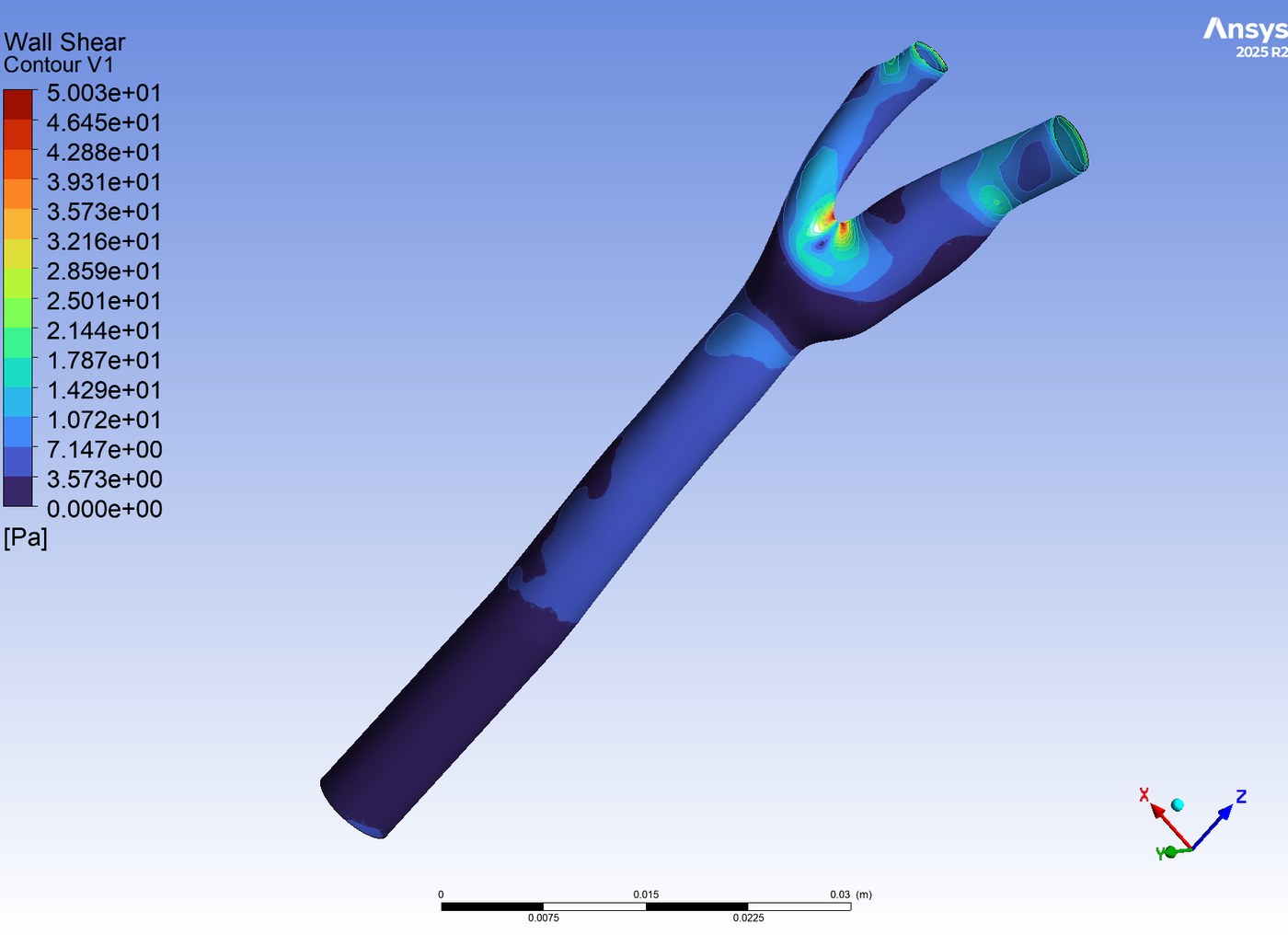
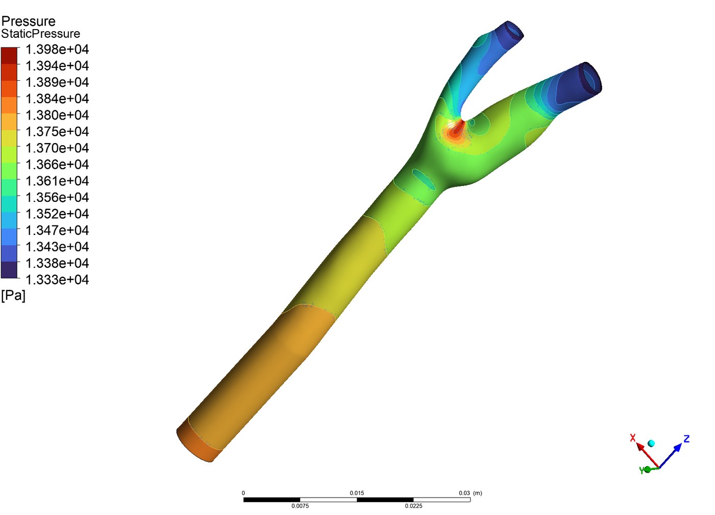
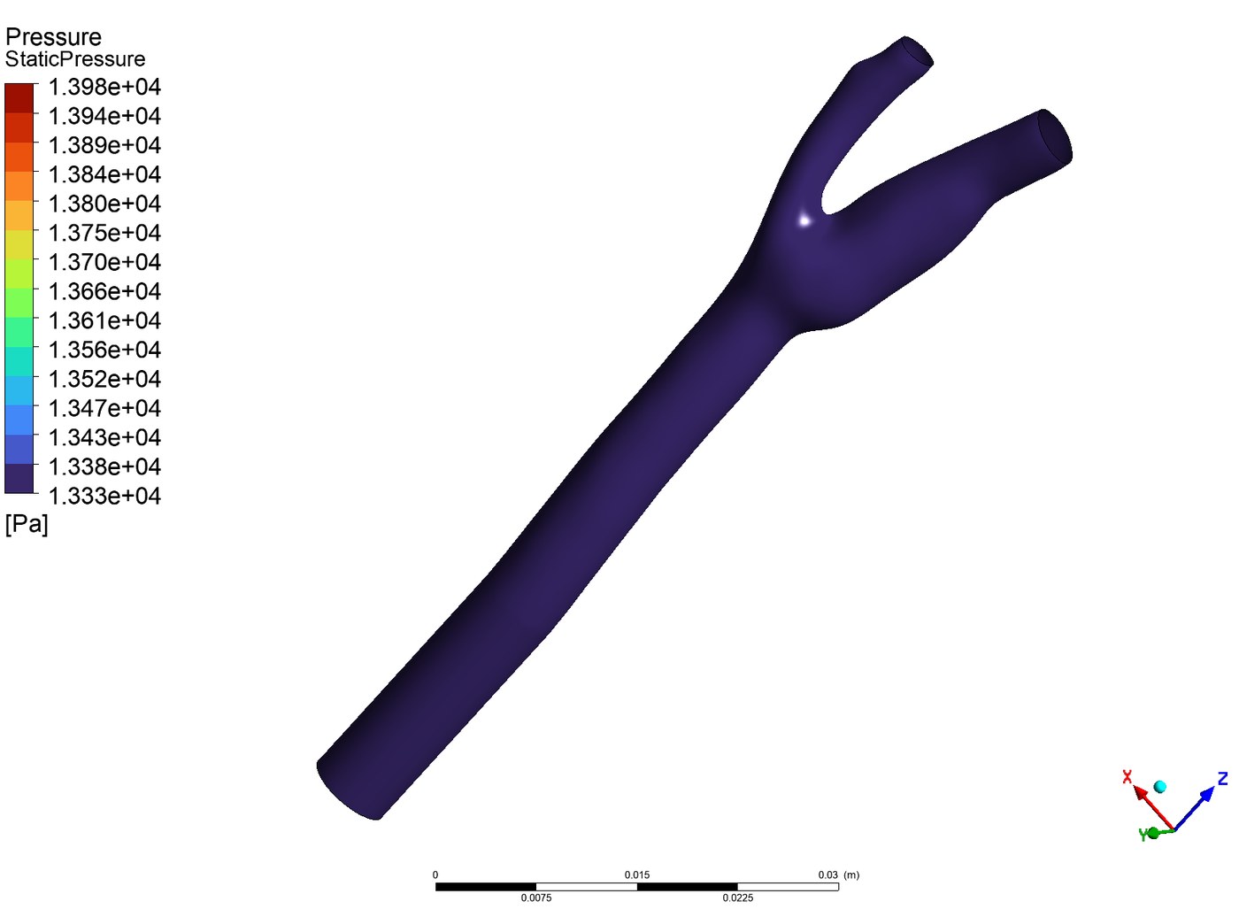
The carotid bifurcation is where the common carotid splits into its internal and external branches, and it is one of the most clinically interesting pieces of vasculature in the body: the geometry produces recirculation and low, oscillating wall shear stress, and those are precisely the conditions plaque forms under. Modeling it well means modeling a real one, so the study starts from an image-based STEP file rather than an idealized tube.
Two preparation steps matter before meshing. The inlet is extended by 20 mm so the velocity profile has room to develop before it reaches the bifurcation, and the surfaces are split into named selections for the inlet, both outlets, and the walls, which is what lets boundary conditions be assigned per face later.
| Mesh stage | Settings |
|---|---|
| Surface mesh | 0.15–0.4 mm, curvature size function, 20° curvature normal angle |
| Boundary layers | 10 layers, smooth-transition, 0.4 transition ratio, 1.2 growth rate, walls only |
| Volume mesh | Poly-hexcore, 3 buffer layers, 2 peel layers, 0.15–0.3 mm cells |
| Quality gate | Minimum orthogonal quality above 0.2, improved further where needed |
The ten inflation layers are the part that earns its cost. Wall shear stress is a velocity gradient evaluated at the wall, so the accuracy of the headline result depends directly on how finely the near-wall region is resolved. A curvature-driven size function does the same job around the bifurcation apex, where the geometry turns sharply.